Launch offer — valid today only

HCAs, Nursing Associates and Returning Nurses: Read a 12-Lead ECG in 8 Steps, Always in the Same Order With Every Wave and Interval Drawn Step by Step!

Rate, rhythm, axis, the PR interval, QRS width, the ST segment, heart blocks, VT and VF: in your notes it's a wall of text. Here every topic fits on one page, with the drawing and what you need to remember. 67 illustrated pages.

SHOW ME THE ILLUSTRATED ECG

🔒 Secure checkout · PDF by email · 30-day guarantee

One topic per pageOne topic per page: the drawing first, the rule in a box
67 illustrated pages8 parts, from the grid and the leads to reading strips on the ward
Up-to-date sourcesResuscitation Council UK ALS guidelines and SCST 12-lead guidance

This Is What the Pages You Will Study Look Like

One topic per page, the drawing that shows you how it's read and the boxes with what you need to know:

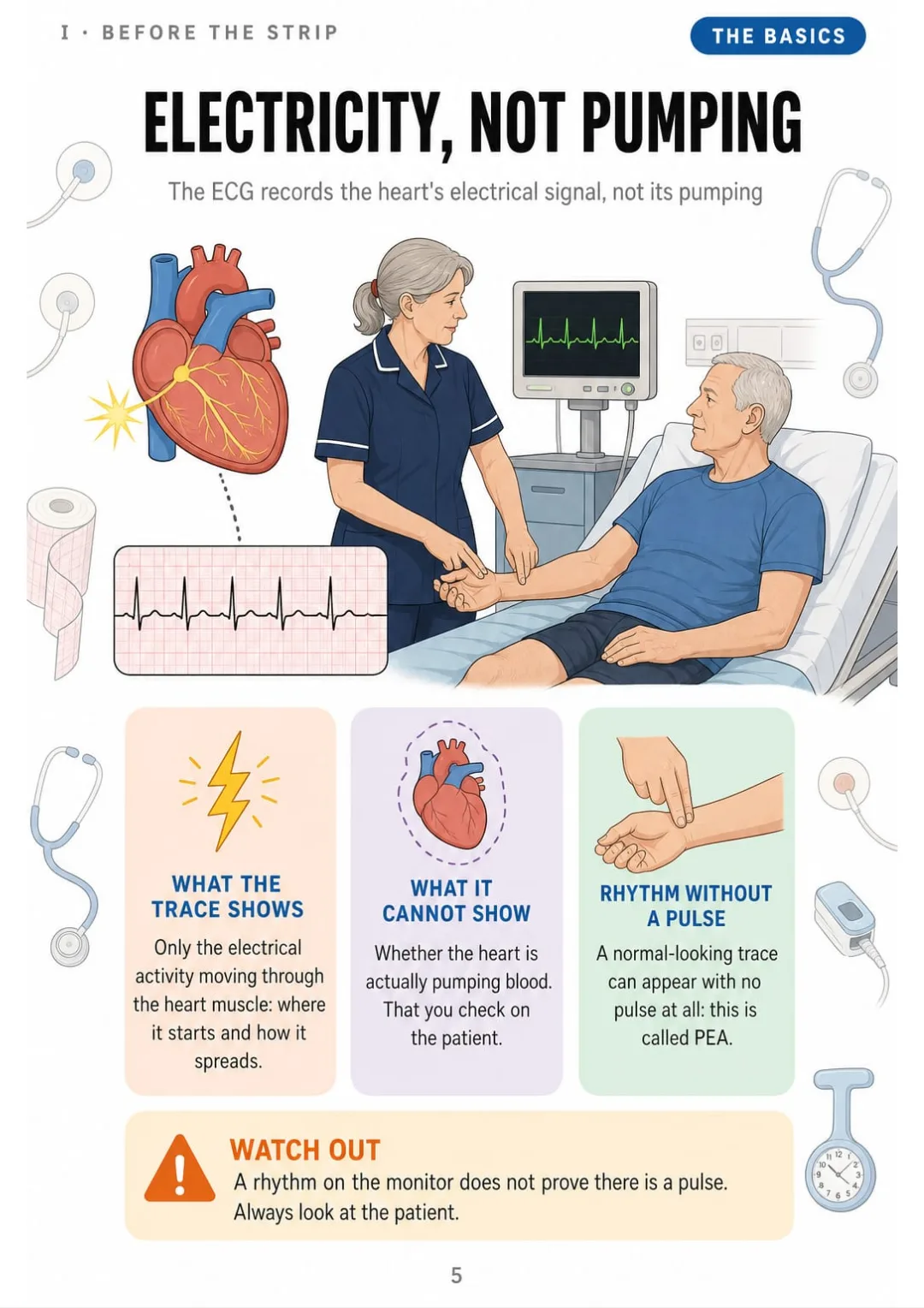
The ECG records electricity, not pumping: a rhythm on screen does not prove a pulse.
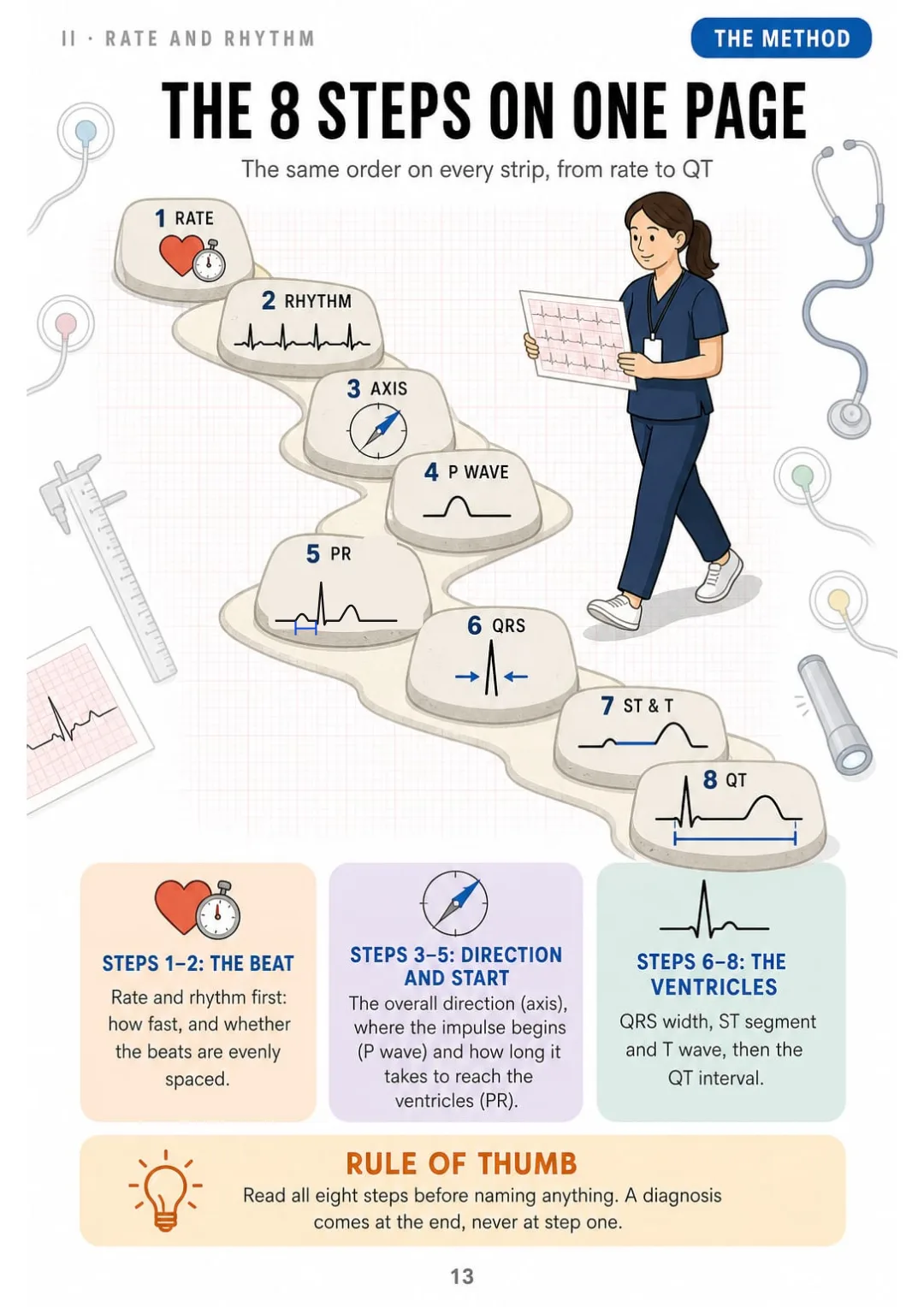
The whole method on one page: rate, rhythm, axis, P, PR, QRS, ST and T, QT.
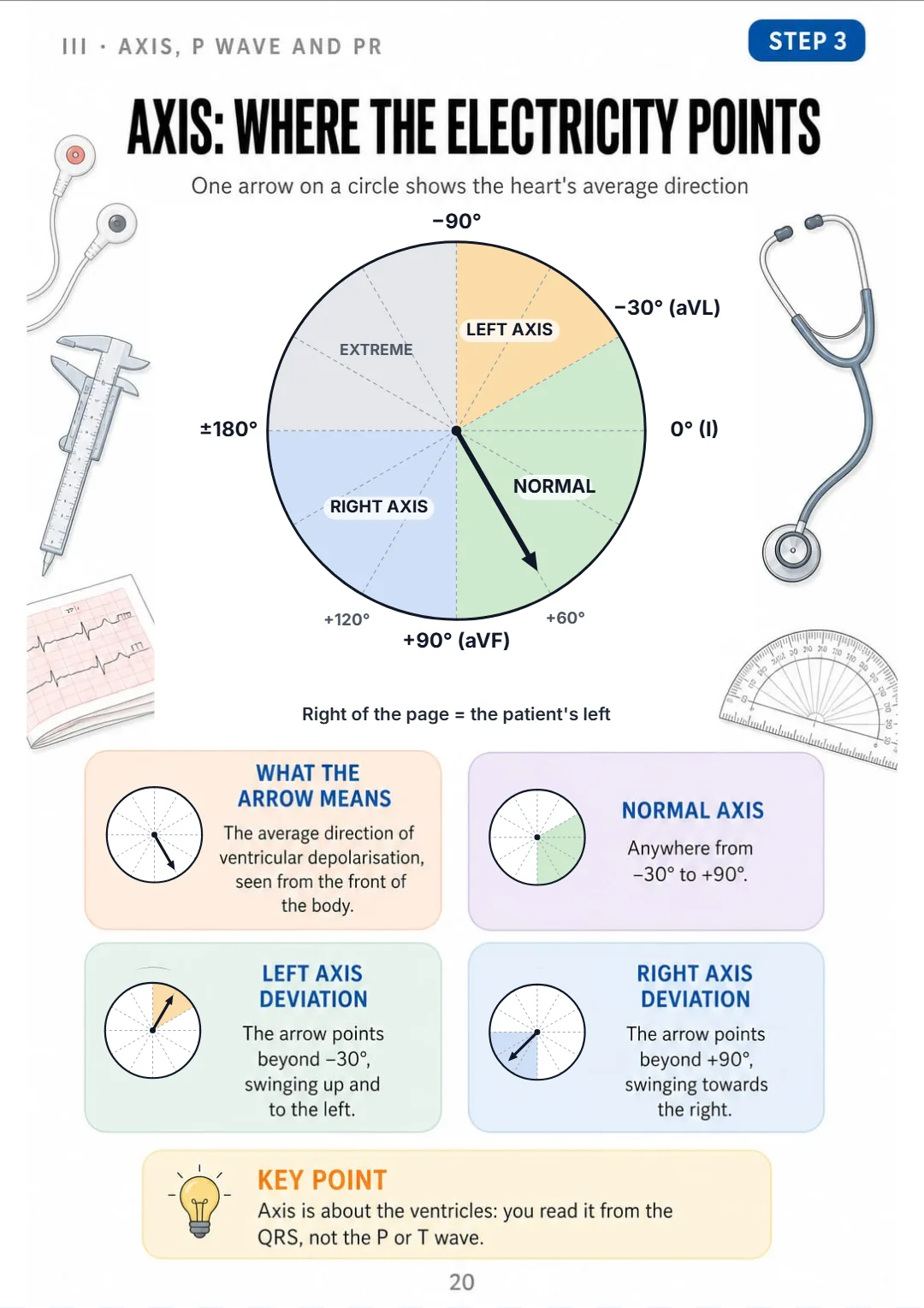
Normal axis −30° to +90°; beyond −30° left, beyond +90° right deviation.
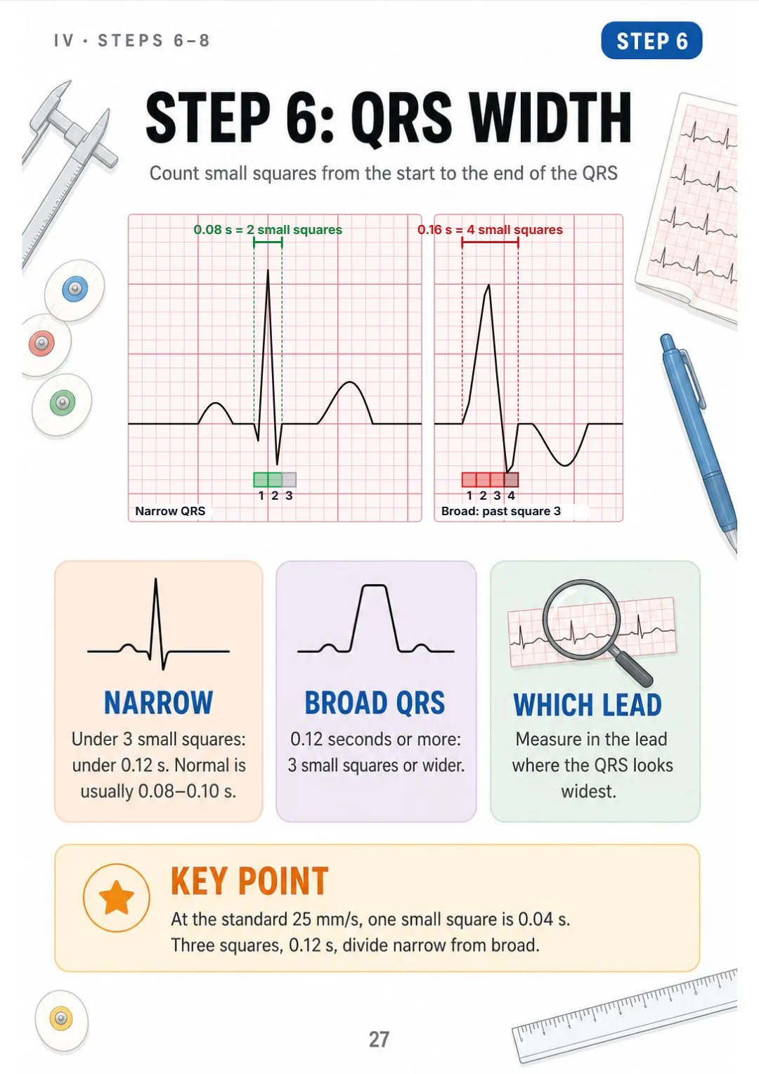
QRS under 0.12 s (3 small squares) is narrow; 0.12 s or more is broad.
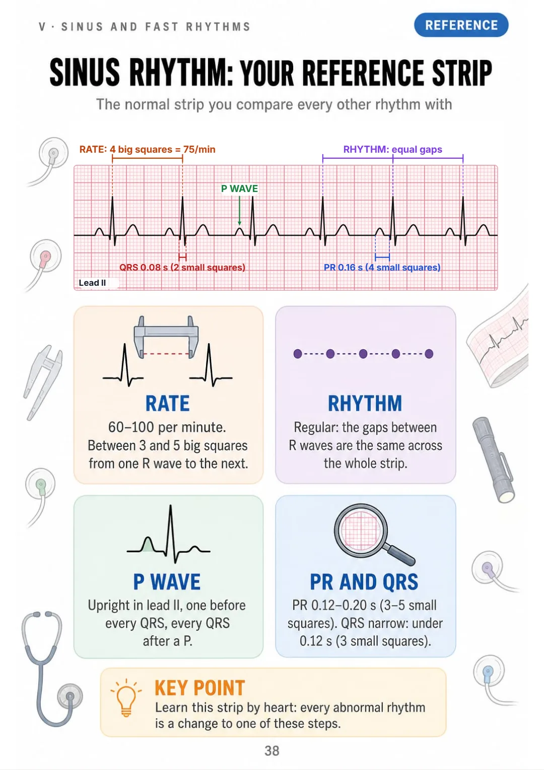
Sinus rhythm: rate 60–100, regular, upright P in II, PR 0.12–0.20 s, narrow QRS.
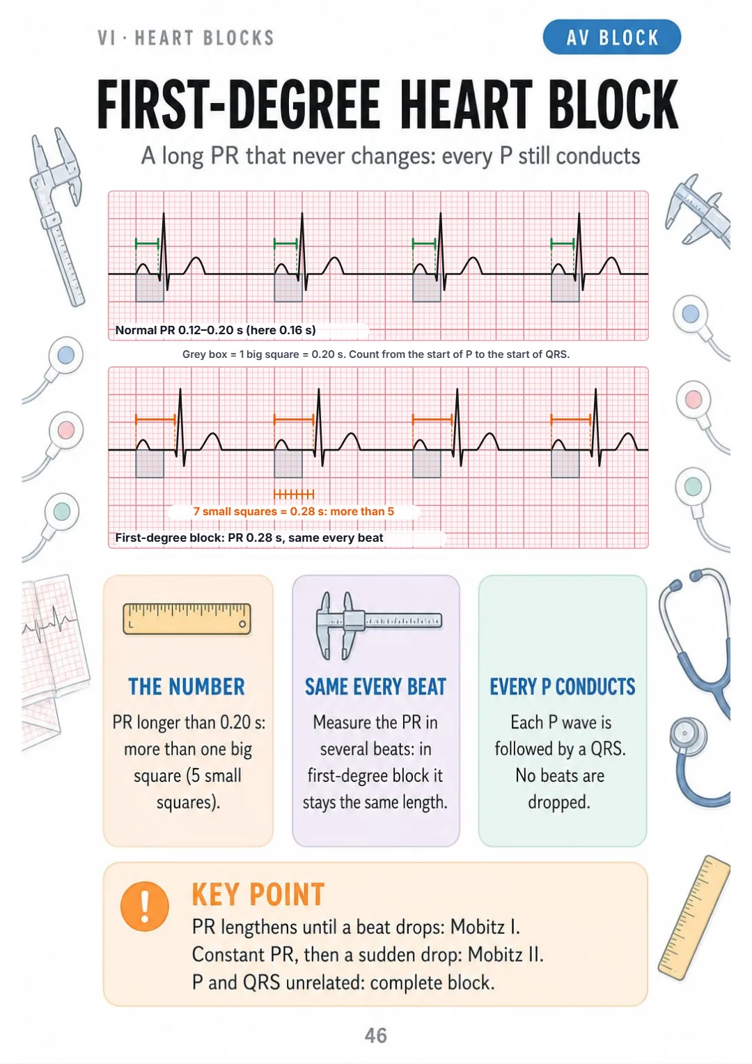
First-degree heart block: PR over 0.20 s, identical every beat, no dropped QRS.
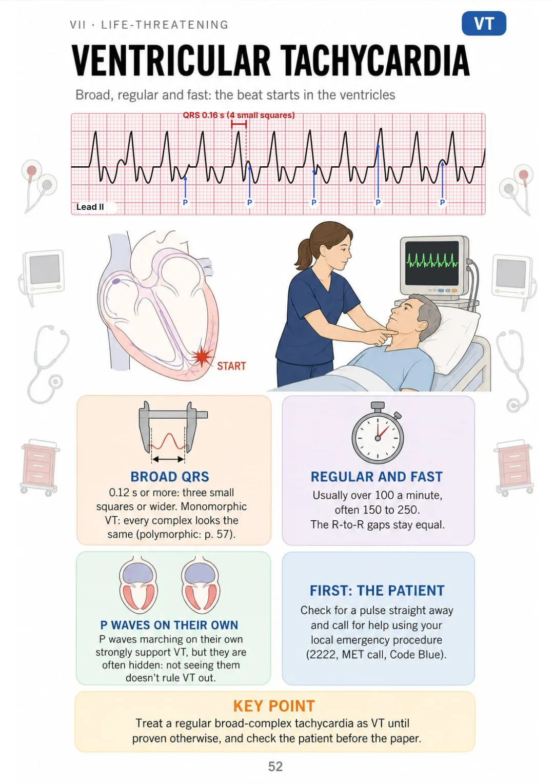
VT: QRS 0.12 s or more, regular, usually over 100 a minute; P waves may march on their own.
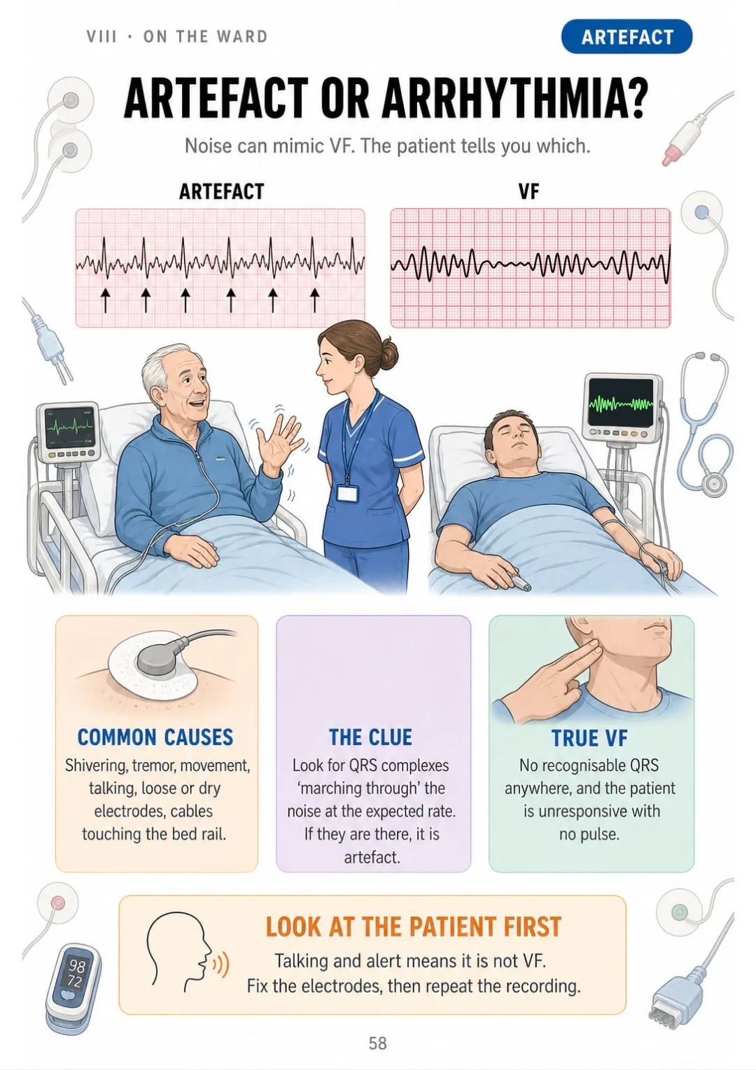
Artefact vs VF: QRS complexes still march through the noise.

Swipe to see more →

These are real pages from the book. The table of contents is the full one: 8 parts and 63 topics.

It's Not That You Can't Remember. It's That Nobody Put It in Order.

You've read the axis chapter three times: degrees, quadrants, a circle full of leads. You close the book...

...and you can't remember which way left axis deviation points. It was just lines of text.

Normal axis −30° to +90°; beyond −30° left, beyond +90° right deviation.

Here the axis is one page: an arrow on a circle, normal from −30° to +90°, and beyond that, left or right deviation, drawn where you can see it.

And on the pages next to it, every step has its own: rate, rhythm, the PR interval, QRS width and the ST segment.

First you understand why it's read that way. Then the order stays with you on its own.

SEE THE DIFFERENCE

Stop memorising ECG rules you can't picture:

THIS
  • Pages and pages of dense text
  • Rhythms without a single drawing
  • In front of the strip, you don't know where to start
Systematic interpretation of the 12-lead electrocardiogram should proceed in a consistent sequence. Initially, the ventricular rate is determined, followed by assessment of rhythm regularity and calculation of the frontal plane QRS axis. Subsequently, P wave morphology and the PR interval are evaluated, after which QRS duration and morphology are examined. Finally, the ST segment, the T wave and the QT interval, corrected for heart rate, are assessed, and all findings are correlated with the clinical presentation of the patient.
FOR THIS
  • Every step drawn
  • One topic per page, with the rules in boxes
  • You remember it because you've seen it
The whole method on one page: rate, rhythm, axis, P, PR, QRS, ST and T, QT.
CONTENTS

Take a Look at the 8 Parts and 63 Topics

The grid and the leads, rate and rhythm, axis and PR, QRS, ST and QT, sinus and fast rhythms, heart blocks, life-threatening rhythms and the ward: 67 pages, all illustrated

IBefore the Strip: Grid, Beat and Leads · pp. 5-12
  1. An ECG Shows Electricity, Not Pumping
  2. The Conduction Pathway, Drawn
  3. One Beat, Labelled
  4. The Grid: Small Square and Big Square
  5. Height and the Calibration Box
  6. 12 Leads, 12 Views
  7. Lead Groups: Which Wall Each Lead Sees
  8. How the Printout Is Laid Out
IISteps 1–2: Rate and Rhythm · pp. 13-19
  1. The 8 Steps on One Page
  2. Rate: the 300 Rule
  3. Rate: the 1500 Rule
  4. Rate: When the Rhythm Is Irregular
  5. Slow, Normal or Fast
  6. Rhythm: Regular or Irregular?
  7. Regularly Irregular vs Irregularly Irregular
IIISteps 3–5: Axis, P Wave and PR · pp. 20-26
  1. Axis: Where the Electricity Points
  2. Axis in 10 Seconds: Leads I and aVF
  3. Step 4: Is There a P Wave?
  4. P Wave Size
  5. Step 5: The PR Interval
  6. Short PR, Long PR
  7. Absent or Odd P Waves
IVSteps 6–8: QRS, ST and T, QT · pp. 27-37
  1. Step 6: QRS Width
  2. Narrow or Broad: Where the Beat Started
  3. R Wave Progression from V1 to V6
  4. Q Waves: Small and Normal, or Significant
  5. Step 7: The ST Segment and the J Point
  6. ST Elevation: Two Leads Next to Each Other
  7. ST Depression
  8. The T Wave: Normal Shapes
  9. Tall, Peaked T Waves
  10. Step 8: The QT Interval
  11. QTc: Why the QT Is Corrected
VSinus and Fast Rhythms · pp. 38-45
  1. Sinus Rhythm: Your Reference Strip
  2. Sinus Bradycardia
  3. Sinus Tachycardia
  4. Sinus Arrhythmia
  5. Atrial Fibrillation
  6. Atrial Flutter
  7. SVT: Narrow, Regular, Fast
  8. Ectopic Beats: Early and Out of Place
VIHeart Blocks and Bundle Branch Blocks · pp. 46-51
  1. First-Degree Heart Block
  2. Second-Degree: Mobitz I (Wenckebach)
  3. Second-Degree: Mobitz II
  4. Third-Degree (Complete) Heart Block
  5. Right Bundle Branch Block
  6. Left Bundle Branch Block
VIILife-Threatening Rhythms · pp. 52-57
  1. Ventricular Tachycardia
  2. Ventricular Fibrillation
  3. Asystole: Check Before You Believe the Line
  4. PEA: a Rhythm Without a Pulse
  5. Shockable and Non-Shockable
  6. Torsades de Pointes
VIIIOn the Ward: Report and Practise · pp. 58-67
  1. Artefact or Arrhythmia?
  2. Swapped Limb Leads
  3. What to Report Straight Away
  4. Saying It Out Loud in 8 Steps
  5. Handing Over an ECG with SBAR
  6. Worked Strip 1: Normal ECG
  7. Worked Strip 2: Atrial Fibrillation
  8. Worked Strip 3: Complete Heart Block
  9. The Most Frequent Mistakes
  10. The 8-Step Pocket Checklist

What If Every Topic Fit on a Single Page?

Imagine this:

Instead of rereading half a page on the QRS without being able to picture it...

...you open the page and find the QRS drawn on the grid: under 3 small squares it's narrow, 3 or more it's broad, and what that tells you about where the beat started.

Sinus rhythm: rate 60–100, regular, upright P in II, PR 0.12–0.20 s, narrow QRS.

And rate, rhythm, axis, heart blocks and VT turn into drawings and tables you can read at a glance.

You stop repeating and start understanding.

That's exactly what The Illustrated ECG does. It isn't ‘just another textbook’. It's 8 parts and 67 illustrated pages, one topic per page, ending with 3 worked strips, the most frequent mistakes and the 8-step pocket checklist.

Because what your brain sees organized, it remembers.

Here Is What You Get with The Illustrated ECG:

Ipp. 5-12

Before the Strip: Grid, Beat and Leads

What the ECG records, the conduction pathway, one beat labelled, the small and big square, the calibration box and the 12 leads with the wall each one sees.

IIpp. 13-19

Steps 1–2: Rate and Rhythm

The 8 steps on one page, the 300 and 1500 rules, rate when the rhythm is irregular, and regular or irregular rhythm.

IIIpp. 20-26

Steps 3–5: Axis, P Wave and PR

Axis with leads I and aVF, the P wave and its size, the PR interval, short and long PR, and absent or odd P waves.

IVpp. 27-37

Steps 6–8: QRS, ST and T, QT

QRS width, R wave progression, Q waves, the ST segment and J point, ST elevation and depression, T waves, and the QT with QTc.

Vpp. 38-45

Sinus and Fast Rhythms

Sinus rhythm as your reference, sinus bradycardia, tachycardia and arrhythmia, AF, atrial flutter, SVT and ectopic beats.

VIpp. 46-51

Heart Blocks and Bundle Branch Blocks

First-degree block, Mobitz I and Mobitz II, complete heart block, and right and left bundle branch block.

VIIpp. 52-57

Life-Threatening Rhythms

VT, VF, asystole, PEA, shockable and non-shockable rhythms, and torsades de pointes.

VIIIpp. 58-67

On the Ward: Report and Practise

Artefact, swapped limb leads, what to report straight away, handing over with SBAR, 3 worked strips, the most frequent mistakes and the pocket checklist.

At the end of the book: 3 worked strips, the most frequent mistakes and the 8-step pocket checklist.

And That Is Not All: 2 More Books, Free

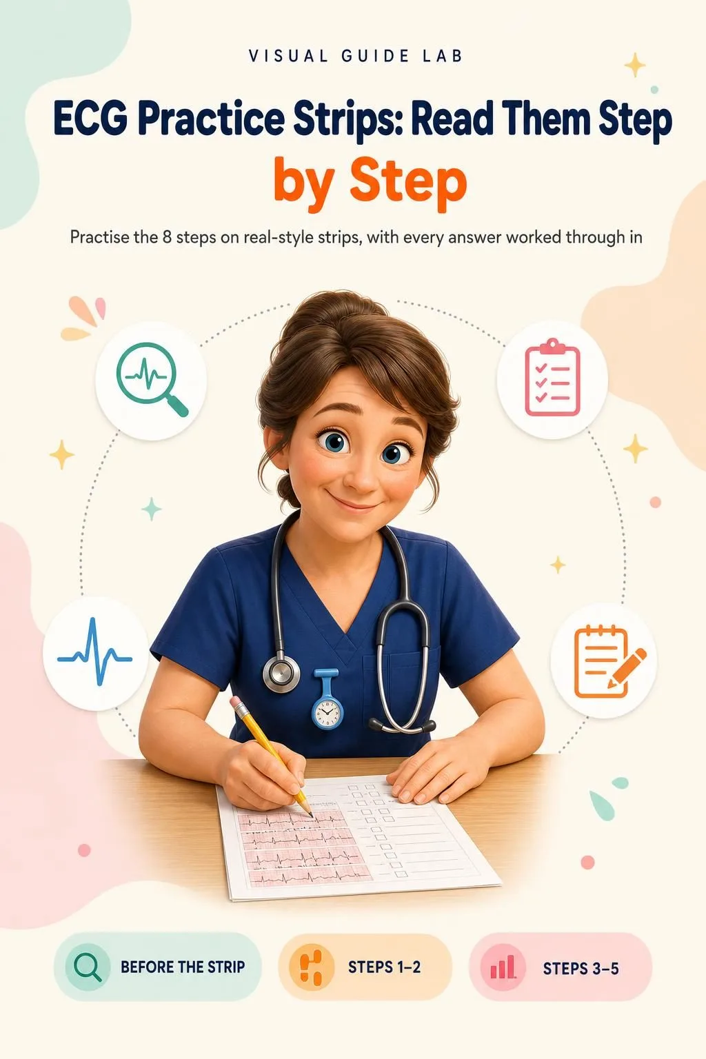
ECG Practice Strips: Read Them Step by Step

BONUS 1: «ECG Practice Strips: Read Them Step by Step»

Value on its own: US$ 17.90

Practise the 8 steps on real-style strips, with every answer worked through in the same order, so the method sticks before your next shift or assessment.

ECG Common Mistakes & Quick-Reference Cards

BONUS 2: «ECG Common Mistakes & Quick-Reference Cards»

Value on its own: US$ 14.90

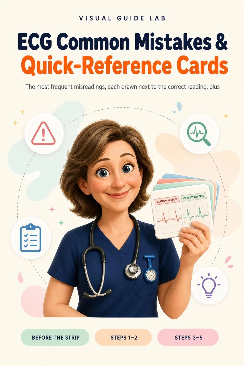
The most frequent misreadings, each drawn next to the correct reading, plus printable pocket cards to check yourself on the ward.

The Illustrated ECG + 2 Free Books

  • The Illustrated ECG: 67 illustrated pagesUS$ 63
  • BONUS 1: ECG Practice Strips: Read Them Step by StepUS$ 17.90
  • BONUS 2: ECG Common Mistakes & Quick-Reference CardsUS$ 14.90
  • High-resolution PDF to printIncluded
Total value US$ 95.80
YOUR PRICE:
US$ 27
You save US$ 68.80
YES, I WANT THE ILLUSTRATED ECG FOR US$ 27

📄 This is a digital PDF book sent to your email: nothing is printed or shipped.

Secure purchaseEncrypted payment30-day guarantee

PDF by email: The Illustrated ECG reaches your inbox as a PDF, and the two free books come by email too. One-time payment, no installments or hidden costs.

Is This Book for You?

You're training as a nursing associate or nurse

You're training as a nursing associate or nurse

The ECG one step per page, to revise between shifts, study days and placement.

Your assessment or placement is coming up

Your assessment or placement is coming up

The whole 8-step method on one page, 3 worked strips and a pocket checklist to check yourself before the day.

You already work on the ward or on the road

You already work on the ward or on the road

A quick visual refresher for HCAs, returning nurses and paramedics: rhythms, heart blocks and what to report straight away.

To Sum Up: Here Is What You Get

US$ 95.80US$ 27
YES, I WANT THE ILLUSTRATED ECG FOR US$ 27

📄 This is a digital PDF book sent to your email: nothing is printed or shipped.

30DAYS
GUARANTEE

30-Day Guarantee

30-day guarantee: tell us what didn't work and we'll refund 100%.

You have 30 days to try The Illustrated ECG and the two bonus books. If it isn't what you needed, email us, tell us what didn't work and we'll refund 100% of what you paid. One-off payment, no subscription, no surprises.

Frequently Asked Questions

How do I receive it?

After you buy, you get an email with The Illustrated ECG as a high-resolution PDF, to read on your phone or print. The two bonus books, ECG Practice Strips: Read Them Step by Step and ECG Common Mistakes & Quick-Reference Cards, also come as PDFs by email.

Is it a monthly or a one-off payment?

A one-off payment of £19.99. No instalments and no subscription. At checkout you'll see some optional extras; you don't need them, and you only pay for what you tick.

Who is it for?

Healthcare assistants who record ECGs and want to read them, nursing associates, student nurses and apprentices, nurses on a Return to Practice programme and paramedics who want a fixed method. It starts from the small square, so you don't need to know ECGs already, and it works as a quick refresher if you do. Have a look at the contents above to see every topic.

What sources is it based on?

It uses the standard reading method taught in the UK and in Australia: standard 25 mm/s paper, the same normal values and the same 8 steps. Arrest and tachycardia content follows Resuscitation Council UK guidance (Adult Advanced Life Support and the adult tachycardia and bradycardia algorithms), which, like ANZCOR's, is based on the same international ILCOR consensus. ECG recording follows the SCST guidelines for a standard 12-lead ECG, and handover is done with SBAR. Always follow your local protocols.

Is it official material?

No. It's independent study material. It isn't published or endorsed by any health service, regulator or resuscitation council (for example the NHS, NMC, HCPC, Resuscitation Council UK, ANZCOR, NMBA or AHPRA), and it doesn't come with a certificate or CPD hours. It's there to help you read ECGs with a fixed method.

Does it replace my course or training?

No. It's study material to revise and practise reading ECGs. It doesn't replace your course, your placement, your employer's protocols or clinical judgement, and it gives no medication or treatment advice.

Are the practice strips and questions from the real assessment?

No. They're written for this book and aren't official questions from any exam or assessment. They're there to train the 8 steps.

Can I print it?

Yes. It's a PDF you can print in full or page by page. The pocket checklist and the quick-reference cards are made to keep to hand.

What if I don't like it?

30-day guarantee: tell us what didn't work and we'll refund 100%.
One Last Thing Before You Decide...

You don't have to reread the same page hoping it sticks this time.

You don't have to memorise rhythms you can't picture.

And you certainly don't have to face the strip without knowing where to start.

There's a simpler way. And it's one click away. Now it's your turn.

One-time payment: US$ 27 · Guarantee: 30 days · PDF by email

YES, I WANT THE ILLUSTRATED ECG FOR US$ 27

📄 This is a digital PDF book sent to your email: nothing is printed or shipped.